Osteoarthritis or OA is the most common cause of joint pain and stiffness. OA occurs with age, or in a joint that was previously injured or overused. There are many conservative treatments for mild OA that may help relieve its painful symptoms.
- Exercise or physical therapy
- Bracing for support
- Anti-inflammatory medication
- Orthotics
- Injection therapy
- Viscosupplementation therapy
Direct Anterior Hip Replacement
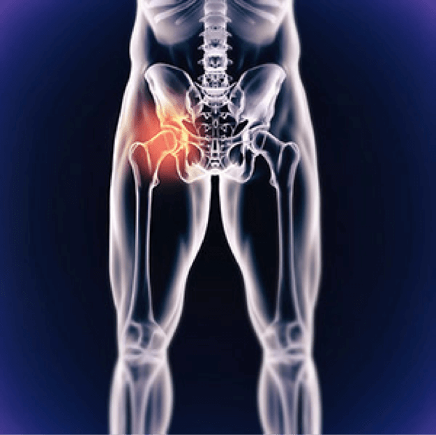 The hip joint is the powerhouse that makes it possible to walk, run and jump.This joint is formed by the union of the top of the femur, the long thigh bone, and the side of the pelvis. The femur has a ball shaped protrusion at the top, which fits snugly into a socket shaped pocket in the pelvis. It is held in place by an intricate system of ligaments and muscles, which provides the locomotive power to move the leg freely in a full circle as well as side-to-side.
Keeping the two bone surfaces from painfully rubbing together is a thick pad of cartilage on both surfaces. The cartilage also acts as a shock
absorber keeping the bone surfaces from crushing together when moving. The cartilage is kept moist by a thin membrane, which provides a watery fluid to lubricate the joint.
The trend for hip replacement surgery is on the rise. The most commonreason for this surgery is arthritis including osteoarthritis, rheumatoid arthritis and traumatic arthritis. Childhood hip diseases can cause problems with the surface of the hip structure later in life even whenthe condition was successfully treated earlier.
Millions of active Americans are seeking help for hip pain because unlike earlier generations, they are not content with modifying or limiting their lives when there are new surgical options available.In the past, patients have used medication, exercise and lifestyle changes as a strategy to postpone surgery. However, when severe pain or joint damage begins to limit your daily activities, a joint replacement is your best option to regain functionality.
Dr. Christopher Vinton performs Direct Anterior Hip Replacements, and is the only surgeon in the local area doing so. This procedure is a technologically advanced and minimally invasive approach to traditional Hip Replacement surgery.
Traditional hip replacement surgery involves making an incision on the side of the hip (lateral approach) or the back of the hip (postero-lateral approach). Both techniques involve detachment of muscles and tendons from the hip to replace the joint. The detachment of these muscles may result in increased pain after surgery, and often prolongs recovery by months or even years. Failure of these muscles to heal after surgery may increase the risk of hip dislocation (the ball and socket separating), which is the leading cause of hip replacement failure. Hip precautions after surgery (no bending greater than 90 degrees, no crossing legs, no excessive rotation) are generally required for traditional hip replacement.
Direct Anterior Hip Replacement is a minimally invasive surgical technique. Using a specialized operating table, called the Hanna table, Dr. Vinton can access the front of the hip. This approach allows the joint to be replaced by moving muscles aside along their natural tissue planes, without detaching any tendons. This approach often results in quicker recovery, less pain, and more normal function after hip replacement. Because the tendons or muscles aren’t detached from the hip during Direct Anterior Hip Replacement, hip precautions are typically not necessary. This allows patients to return to normal daily activities shortly after surgery with a reduced risk of dislocation.
For more information, read about Anterior vs. Posterior Hip Replacement Surgeries.
Total Knee Replacement
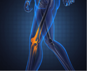 The knee is one of the most complex joints in the body because it is heavily responsible for both the horizontal and vertical motion in the body.
The knee consists of three primary parts:
- Lower part of the thighbone (femur)
- Upper end of the shinbone (tibia)
- Kneecap (patella)
These three bones form a junction. The end of each bone is covered with a substance called articular cartilage. This articular cartilage is smooth, providing a surface along which the bones can easily move against each other. The thighbone and shinbone are held together with a series of ligaments, and the kneecap is placed in front of this junction for protection.
When all components are working in harmony, the knee is a remarkable feat of engineering. When disease or injury disrupts its operation, the resultant pain and muscle weakness can be debilitating. Unfortunately, the knee is prone to arthritis and other conditions that reduce its ability to move smoothly and painlessly. If a knee replacement is necessary, Drs. Lahey and Vinton have ample experience in total knee replacement.
Partial Knee Replacement
 Dr. Philip Lahey IV performs both total and partial knee replacement. In many cases, osteoarthritis only occurs in one compartment (portion) of the knee. A Partial Knee Replacement, also known as a unicompartmental knee replacement, will resurface only the damaged side of the knee, preserving the normal, undamaged cartilage. This allows for a smaller incision. The four natural ligaments of the knee also remain intact. Other advantages of a partial knee include less pain, a faster recovery, and a more natural motion. The Oxford Partial Knee (Zimmer Biomet) is the most widely used and clinically proven partial knee in the world. Long-term clinical results on the Oxford Partial Knee demonstrate that 94% of implants are still functioning well at 15 years, and 91% are functioning well at 20 years.
For more information: http://www.biomet.com/patients/oxford.cfm